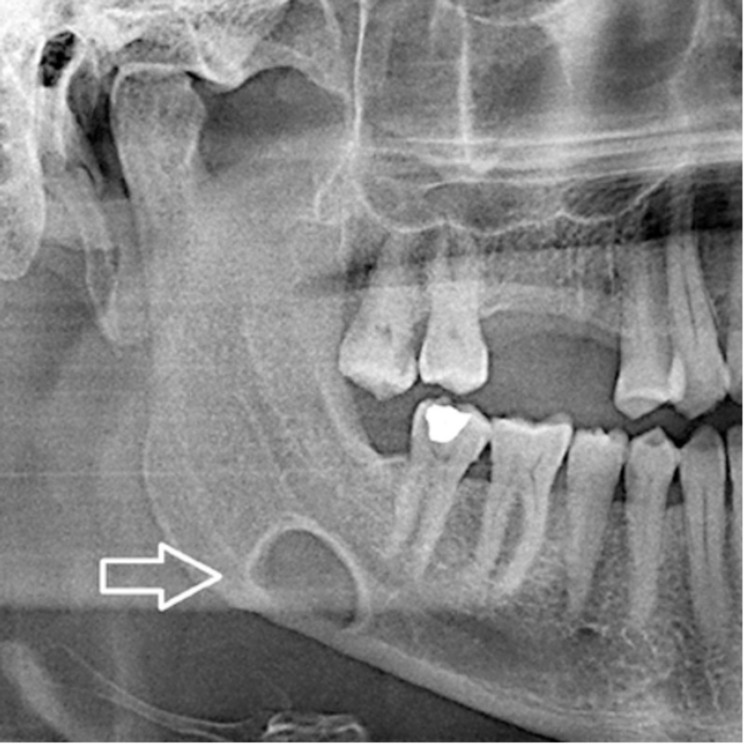
“What Is The Difference Between An Aneurysmal Bone Cyst And Stafne Bone Defect?”
6.4.24 Quick Review #141 - #pathology #oralpathology #doctorgallagher #oralsurgery #oralsurgeon #dentist #dentistry #dental An aneurysmal bone cyst (ABC) and a Stafne bone defect (SBD) are both bone lesions but they differ significantly in their nature, location, and clinical implications: Aneurysmal Bone Cyst (ABC) 1. Nature: - ABCs are benign but potentially aggressive bone lesions. - They are characterized by blood-filled spaces separated by connective tissue septa containing osteoid tissue and osteoclast giant cells. 2. Location: - ABCs can occur in any bone but are most commonly found in the long bones and the vertebrae. - They are relatively rare in the jaws but can still occur there. 3. Etiology: - The exact cause is unknown, but they are thought to arise secondary to trauma, a vascular malformation, or as a secondary lesion to other bone tumors. 4. Clinical Features: - Symptoms often include pain, swelling, and potential pathological fractures. - They can cause significant bone expansion and destruction. 5. Radiographic Appearance: - ABCs typically appear as expansile, lytic lesions with a "soap-bubble" or "honeycomb" appearance. - They often have well-defined margins. 6. Treatment: - Treatment often involves surgical curettage, sometimes combined with bone grafting. - Recurrence can occur, so follow-up is necessary. Stafne Bone Defect (SBD) 1. Nature: - SBDs, also known as static bone cysts or Stafne cysts, are asymptomatic, non-progressive bone defects. - They are considered developmental anomalies rather than true cysts or tumors. 2. Location: - SBDs are most commonly located in the posterior mandible, typically below the mandibular canal near the angle of the jaw. - They are rarely found in other locations. 3. Etiology: - SBDs are believed to be caused by ectopic salivary gland tissue entrapped during the development of the mandible. 4. Clinical Features: - They are usually discovered incidentally on radiographs taken for other reasons. - Patients are typically asymptomatic. 5. Radiographic Appearance: - SBDs appear as well-defined, round or oval radiolucencies with a corticated border. - They are located below the inferior alveolar nerve canal. 6. Treatment: - No treatment is required as they are benign and non-progressive. - Periodic radiographic monitoring may be done to confirm stability. References: 1. Castilho, E. C., Vieira, E. M., & Cavalcanti, H. G. (2018). Stafne bone cavity containing ectopic parotid gland. Brazilian Journal of Otorhinolaryngology, 84(5), 676-679. 2. Miloro, M., Ghali, G. E., Larsen, P. E., & Waite, P. (Eds.). (2022). Peterson’s Principles of Oral and Maxillofacial Surgery (4th ed.). Springer. 3. Abubaker, A.O., Lam, D., & Benson, K. (2016). OMFS Secrets (3rd ed.). Elsevier. 4. ChatGPT. 2024. #podcast #podcasts #dentalpodcast #dentalpodcasts #doctorgallagherpodcast #doctorgallagherspodcast
You May Also Like

Playlist Culture G : les podcasts pour apprendre chaque jour
Acast France

Un bonbon sur la langue
RTL

Neville Goddard - Classic Radio
Neville Goddard

Neville Goddard - Old Time Radio
Neville Goddard

Chat GPT Collective Consciousness
Sol Good Network

The Mnemonic Memory Podcast
Peter Jans

Estoicismo para Ganadores
Estoicismo Para Ganadores

【本・再放送】英語の語源が身につくラジオ (heldio)
堀田隆一

Motivational Speeches
Motivationly

Fluent Fiction - Catalan
FluentFiction.org
Playlist Culture G : les podcasts pour apprendre chaque jour
Acast France
Un bonbon sur la langue
RTL
Neville Goddard - Classic Radio
Neville Goddard
Neville Goddard - Old Time Radio
Neville Goddard
Chat GPT Collective Consciousness
Sol Good Network
The Mnemonic Memory Podcast
Peter Jans
Estoicismo para Ganadores
Estoicismo Para Ganadores
【本・再放送】英語の語源が身につくラジオ (heldio)
堀田隆一
Motivational Speeches
Motivationly
Fluent Fiction - Catalan
FluentFiction.org
Community discussion