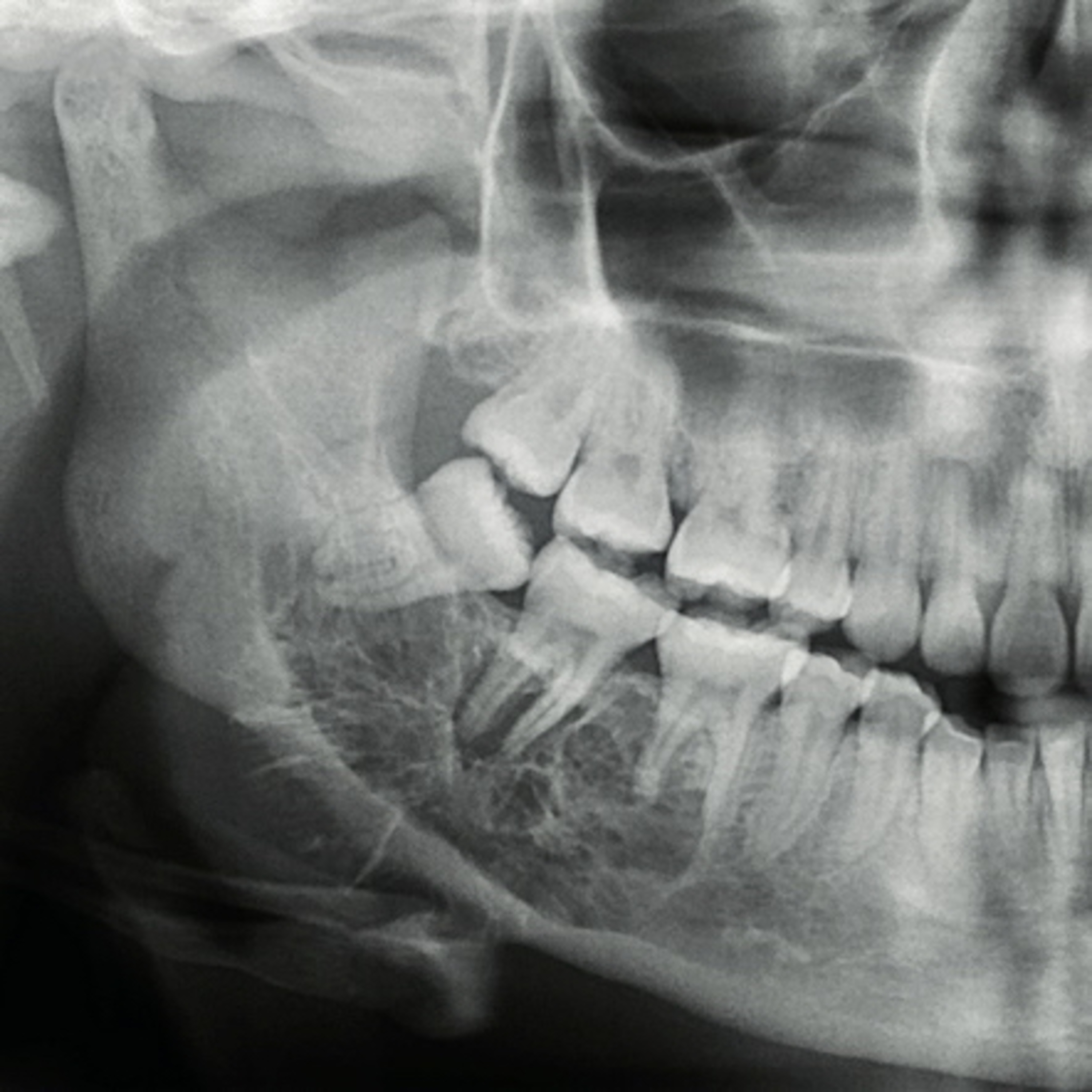
“What are the Key Radiographic Features of the Odontogenic Myxoma?”
2.22.24 Quick Review #111 - #pathology #oralpathology #doctorgallagher #oralsurgery #oralsurgeon #dentist #dentistry #dental Odontogenic myxoma is a benign, but locally aggressive tumor with distinctive radiographic features that help in its diagnosis: 1. Multilocular Radiolucency: One of the hallmark signs of odontogenic myxoma is its multilocular appearance, often described as resembling a “step-ladder” appearance, or “soap bubbles” or a “honeycomb”. This feature is characterized by multiple radiolucent areas separated by thin septa, giving the lesion a multicystic appearance. 2. Well-defined Margins: Although aggressive, odontogenic myxomas often have well-defined margins. This clarity, however, does not imply a less aggressive nature, as the lesion can still infiltrate surrounding bone tissues. 3. Non-corticated Borders: Unlike some other lesions that have a dense, sclerotic border (corticated), odontogenic myxomas typically present with non-corticated, somewhat diffuse borders, making it challenging to determine the lesion’s full extent. 4. Scalloping between the Roots: The lesion may exhibit a scalloped pattern extending between the roots of adjacent teeth. Unlike some aggressive tumors, odontogenic myxoma tends to displace rather than resorb the roots, maintaining the integrity of the tooth structures it surrounds. 5. Displacement of Adjacent Structures: Odontogenic myxomas can cause displacement of teeth and expansion of the jawbones. The teeth adjacent to or within the lesion are often moved apart, but significant root resorption is uncommon, distinguishing it from more invasive pathologies. 6. Size and Expansion: These tumors can grow to be quite large and often cause significant expansion of the jawbones. This expansion is usually buccolingual, leading to cortical thinning and potential perforation, visible on radiographs. 7. Rarity of Calcification: Unlike some other tumors, odontogenic myxomas rarely exhibit internal calcifications within the lesion. The absence of calcified structures within the radiolucent areas can help differentiate it from other mixed radiolucent-radiopaque lesions. References: 1. ResearchGate. (2017). Odontogenic myxoma. Characteristic radiolucency with fine internal opaque trabeculations of the right posterior mandible. Retrieved from https://www.researchgate.net/figure/ 2. Martínez-Pérez, D., & Varela-Morales, M. (2001). Conservative treatment of odontogenic myxoma: report of two cases. Oral Surgery, Oral Medicine, Oral Pathology, Oral Radiology, and Endodontics, 91(5), 569-573. 3. Lo Muzio, L., Nocini, P., Favia, G., Procaccini, M., & Testa, N. F. (1996). Odontogenic myxoma: report of two cases. Clinical Oral Investigations, 1(1), 54-58. 4. ChatGPT. 2024.
You May Also Like

Shimon Davis
Shimon Davis

Choses à Savoir - Culture générale
Choses à Savoir

EconTalk
Russ Roberts

Jabulujule 9 -12
ukhozi fm

Lana Blakely
Lana Blakely

Financial Coaching for Women: How To Budget, Manage Money, Pay Off Debt, Save Money, Paycheck Plans
Vanessa and Shana | Budget Besties | Bougie On A Budget | Dave Ramsey Fans

Neville Goddard - Old Time Radio
Neville Goddard

Neville Goddard Daily
Neville Goddard

Chat GPT Questions
Sol Good Network

Chat GPT Collective Consciousness
Sol Good Network
Shimon Davis
Shimon Davis
Choses à Savoir - Culture générale
Choses à Savoir
EconTalk
Russ Roberts
Jabulujule 9 -12
ukhozi fm
Lana Blakely
Lana Blakely
Financial Coaching for Women: How To Budget, Manage Money, Pay Off Debt, Save Money, Paycheck Plans
Vanessa and Shana | Budget Besties | Bougie On A Budget | Dave Ramsey Fans
Neville Goddard - Old Time Radio
Neville Goddard
Neville Goddard Daily
Neville Goddard
Chat GPT Questions
Sol Good Network
Chat GPT Collective Consciousness
Sol Good Network
Community discussion