📝 What are the Key Differences Between Proliferative Verrucous Leukoplakia & Verrucous Carcinoma?
- 10.28.24
Quick Review #250 - #pathology #oralpathology #doctorgallagher #oralsurgery #oralsurgeon #dentist #dentistry #dental #oralcancer #cancer
Clinical:
• Proliferative Verrucous Leukoplakia (PVL):
• PVL is a progressive, multifocal, and recurrent form of oral leukoplakia.
• It often presents as a slow-growing white patch with a high likelihood of recurrence and malignant transformation.
• PVL primarily affects older adults, with a female predilection.
• Over time, PVL lesions tend to become more aggressive, transforming into verrucous carcinoma or squamous cell carcinoma.
• Verrucous Carcinoma (VC):
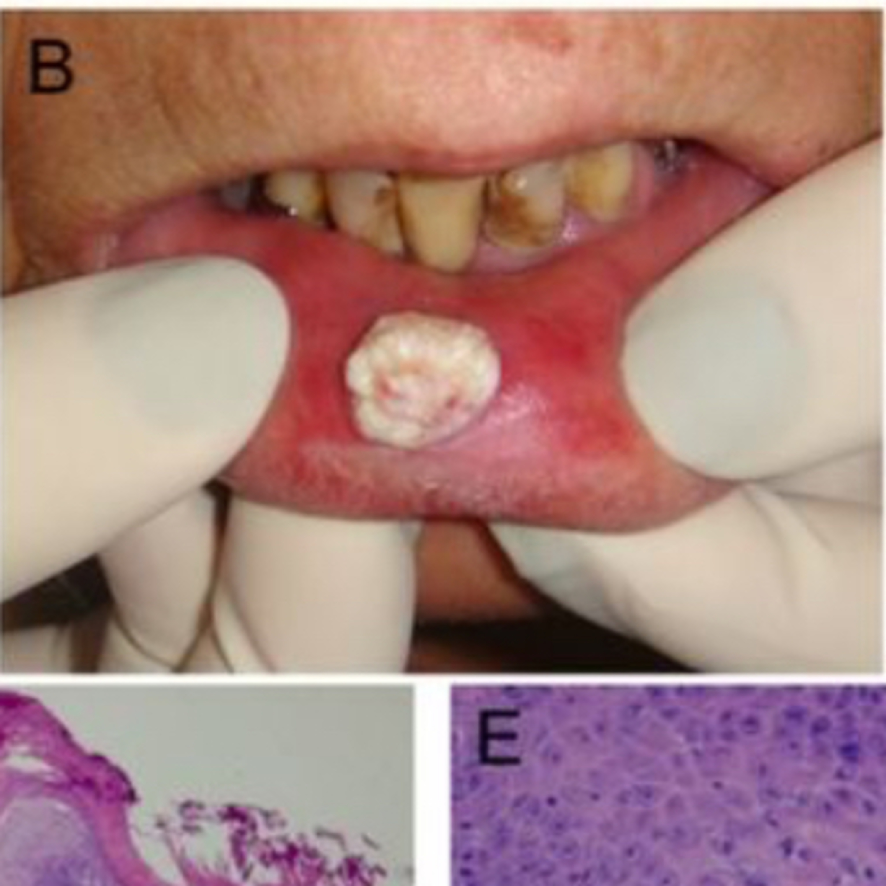
• VC is a well-differentiated variant of squamous cell carcinoma.
• It manifests as an exophytic, warty, or cauliflower-like growth with minimal tendency for metastasis but can invade local tissues.
• Typically affects older adults and is often associated with tobacco use, especially smokeless forms.
Radiographic:
• PVL lesions are often limited as it primarily affects the mucosa; however, it can occasionally show mild bone involvement in advanced stages, particularly if there’s malignant transformation.
• VC may show evidence of local invasion in advanced cases, such as bone erosion, especially if it invades deeply into surrounding tissues. Radiographs can help assess the depth and extent of invasion in cases with substantial growth.
Histopathological:
• PVL is characterized by hyperkeratosis and dysplasia. Early PVL lesions show features of hyperplastic epithelium and may lack overt dysplasia.
• As PVL progresses, it can display various degrees of dysplasia, leading to carcinoma in situ or invasive carcinoma.
• PVL typically has a slow, progressive increase in atypia, often necessitating longitudinal histopathological evaluation due to its transformation risk.
• VC histopathologically demonstrates a thickened, verrucous (warty) epithelial surface with minimal cellular atypia.
• It has a “pushing” pattern of invasion rather than the usual infiltrative invasion seen in other carcinomas, with broad, bulbous rete ridges that extend into the underlying connective tissue.
• VC generally lacks the significant dysplasia seen in PVL, although it may coexist with areas of conventional squamous cell carcinoma, particularly in advanced or long-standing cases.
References:
1. Camisasca, D. R. (n.d.). Oral verrucous carcinoma of the lip mucosa: Typical clinical presentation showing an indolent sessile nodule with a white verrucous surface [Figure]. ResearchGate
2. Neville, B. W., Damm, D. D., Allen, C. M., & Chi, A. C. (2015). Oral and maxillofacial pathology (4th ed.). Elsevier
3. ChatGPT.2024
#podcast #podcasts #dentalpodcast #dentalpodcasts #doctorgallagherpodcast #doctorgallagherspodcast #doctor #dentistry #oralsurgery #dental #viral #dentalschool #dentalstudent #omfs #surgeon #doctorlife #dentistlife #residency #oralsurgeon #dentist #doctorgallagher
You May Also Like

Law School Admissions Unplugged Podcast: Personal Statements, Application Essays, Scholarships, LSAT Prep, and More…
Steve Schwartz

LSAT Unplugged + Law School Admissions Podcast
Steve Schwartz

Ethan Brings You the World
Ethan Boronski

sarah marie anderson's podcast
sarah marie anderson

Shimon Davis
Shimon Davis

Feeling Things with Amy & Kat
Nashville Podcast Network

今を感じ,積重ね,今律・今立
岩田良太

SHIR HADASH
Shir Hadash

CANITY
CANITY

Teach Me to Fish "Helping Leaders better understand what drives People"
Matt Leclair
Law School Admissions Unplugged Podcast: Personal Statements, Application Essays, Scholarships, LSAT Prep, and More…
Steve Schwartz
LSAT Unplugged + Law School Admissions Podcast
Steve Schwartz
Ethan Brings You the World
Ethan Boronski
sarah marie anderson's podcast
sarah marie anderson
Shimon Davis
Shimon Davis
Feeling Things with Amy & Kat
Nashville Podcast Network
今を感じ,積重ね,今律・今立
岩田良太
SHIR HADASH
Shir Hadash
CANITY
CANITY
Teach Me to Fish "Helping Leaders better understand what drives People"
Matt Leclair
Community discussion