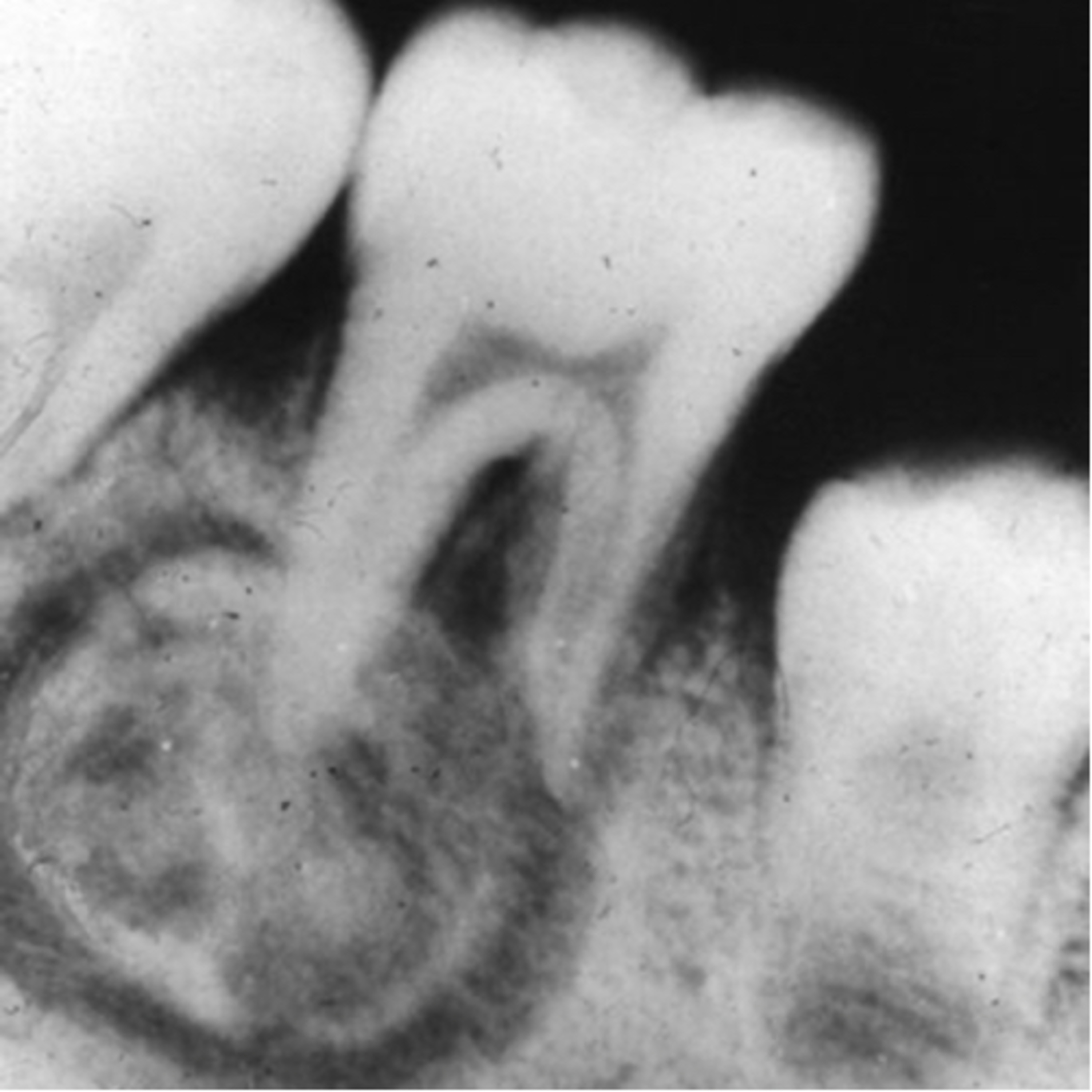
“What are the Differences Between the Osteoma, Cementoblstoma, and Cemento-Ossifying Fibroma?”
7.16.24 Quick Review #180 - #pathology #oralpathology #doctorgallagher #oralsurgery #oralsurgeon #dentist #dentistry #dental Osteoma - benign tumors composed of mature, compact, or cancellous bone. Characteristics: • Growth: Slow-growing. • Location: Commonly found in the craniofacial region, especially the paranasal sinuses, mandible, and skull. • Radiographic Appearance: Radiopaque mass with a well-defined border; it can be sessile or pedunculated. • Symptoms: Often asymptomatic but can cause swelling or asymmetry if large. Histopathology: • Composed of dense, mature bone with little marrow spaces. Clinical Significance: • Generally, no malignant transformation. • May be associated with Gardner’s syndrome, which includes multiple osteomas, colorectal polyps, and soft tissue tumors. Cementoblastoma - benign tumors of cementum-producing cells (cementoblasts). Characteristics: • Growth: Slow-growing but can be aggressive. • Location: Most commonly associated with the roots of mandibular molars and premolars. • Radiographic Appearance: Well-defined radiopaque mass attached to the root of a tooth, often with a surrounding radiolucent halo. • Symptoms: Pain and swelling are common due to the tumor’s pressure on the surrounding bone and nerves. Histopathology: • Consists of sheets of cementum-like material with entrapped cementoblasts and cementocytes. Clinical Significance: • Can cause root resorption. • Treatment often involves surgical removal of the tumor and associated tooth. Cemento-Ossifying Fibroma (COF) - a benign fibro-osseous lesion composed of fibrous tissue, bone, and cementum-like material. Characteristics: • Growth: Slow-growing but can cause significant expansion. • Location: Commonly affects the mandible, especially the premolar and molar regions. • Radiographic Appearance: Mixed radiolucent-radiopaque lesion with a well-defined border; early stages may appear more radiolucent, becoming more radiopaque as it matures. • Symptoms: May cause facial asymmetry, swelling, and displacement of teeth. Histopathology: • Composed of fibrous connective tissue with varying amounts of calcified bone and cementum-like material. Clinical Significance: • Treated with surgical excision. • Recurrence is uncommon but can occur if not completely removed. References: 1. Dr. G’s Toothpix. (n.d.). Cementoblastoma. Retrieved July 16, 2024, from https://lnkd.in/eK2eDCxR 2. Barnes, L., Eveson, J. W., Reichart, P., & Sidransky, D. (2005). World Health Organization Classification of Tumours: Pathology and Genetics of Head and Neck Tumours. IARC Press. 3. Neville, B. W., Damm, D. D., Allen, C. M., & Chi, A. C. (2016). Oral and Maxillofacial Pathology (4th ed.). Elsevier. 4. ChatGPT. 2024. - #podcast #podcasts #dentalpodcast #dentalpodcasts #doctorgallagherpodcast #doctorgallagherspodcast #doctor #dentistry #oralsurgery #dental #viral #dentalschool #dentalstudent #omfs #surgeon #doctorlife #dentistlife #residency #oralsurgeon #dentist #doctorgallagher
You May Also Like
Kabbalah Media | mp3 #kab_ita
Bnei Baruch Kabbalah Education and Research Institute, Бней Барух – Ассоциация "Каббала Ла-Ам"

Make Your Damn Bed
Julie Merica
קבלה מדיה | mp4 #kab_heb
Bnei Baruch Kabbalah Education and Research Institute, Бней Барух – Ассоциация "Каббала Ла-Ам"
Cabalá: Lecciones Diarias | mp4 #kab_spa
Bnei Baruch Kabbalah Education and Research Institute, Бней Барух – Ассоциация "Каббала Ла-Ам"

Feeling Things with Amy & Kat
Nashville Podcast Network

Hamza Selfimprovement
Adonis Gang
Kabbalah Media | mp4 #kab_ger
Bnei Baruch Kabbalah Education and Research Institute, Бней Барух – Ассоциация "Каббала Ла-Ам"
Kabbalah Media | mp3 #kab_fre
Bnei Baruch Kabbalah Education and Research Institute, Бней Барух – Ассоциация "Каббала Ла-Ам"
Kabbalah Media | mp4 #kab_fre
Bnei Baruch Kabbalah Education and Research Institute, Бней Барух – Ассоциация "Каббала Ла-Ам"

Yakın İlişkiler
Podbee Media
Community discussion