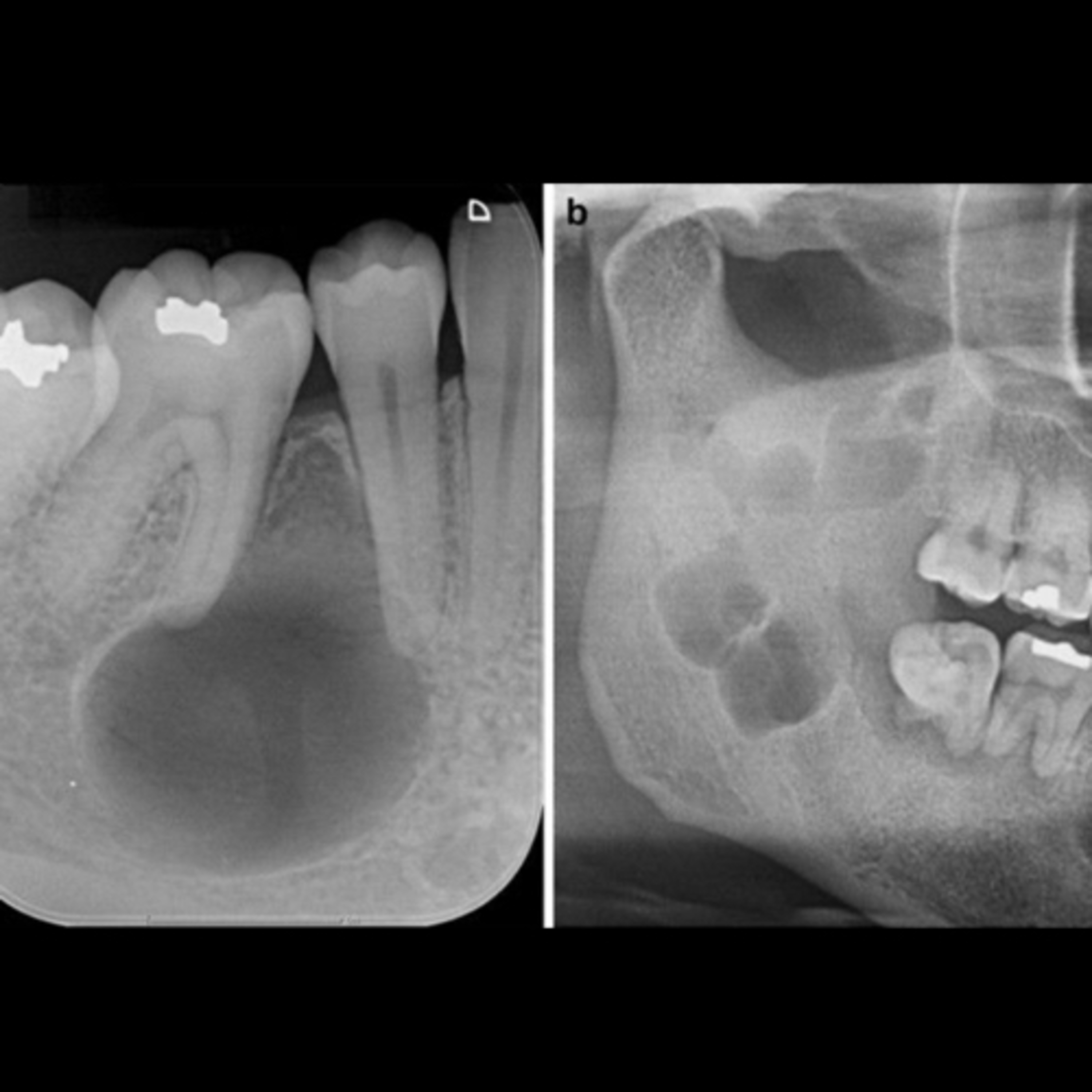
“What Are The Differences Between An Ameloblastoma & An Odontogenic Keratocyst?”
6.20.24 Quick Review #154 - #pathology #oralpathology #doctorgallagher #oralsurgery #oralsurgeon #dentist #dentistry #dental Clinical Features: Ameloblastoma: • Slow-growing, benign but locally aggressive tumor. • Commonly occurs in the mandible, especially the molar-ramus area. • Painless swelling or expansion of the jaw. • Can cause tooth displacement and root resorption. Odontogenic Keratocyst (OKC): • Also known as keratocystic odontogenic tumor. • Aggressive behavior with a high recurrence rate. • Often asymptomatic, but may present as swelling or pain if infected. • Commonly found in the posterior mandible, often associated with an impacted tooth. Radiographic Features: Ameloblastoma: • Multilocular (soap bubble or honeycomb appearance) radiolucency. • May present as unilocular radiolucency. • Well-defined and corticated margins. • Possible cortical bone expansion and perforation. Odontogenic Keratocyst: • Unilocular or multilocular radiolucency. • Smooth, corticated margins. • Tendency to grow along the length of the bone without significant expansion. • Can be associated with an unerupted tooth. Histopathological Features: Ameloblastoma: • Follicular pattern: Islands of odontogenic epithelium with peripheral palisading and reverse polarization. • Plexiform pattern: Interconnected strands or cords of odontogenic epithelium. • Stroma is typically fibrous and may contain cystic changes. Odontogenic Keratocyst: • Lined by a uniform layer of parakeratinized stratified squamous epithelium. • Corrugated surface of the epithelial lining. • Basal layer is palisaded and hyperchromatic. • Cyst wall often contains daughter cysts or epithelial rests. Treatment: Ameloblastoma: • Surgical resection with wide margins to prevent recurrence. • Options include enucleation with curettage, marginal resection, or segmental resection depending on the size and location. • Close long-term follow-up due to potential for recurrence. Odontogenic Keratocyst: • Enucleation with or without adjunctive therapies (Carnoy’s solution, liquid nitrogen cryotherapy). • Marsupialization or decompression followed by enucleation for large lesions. • Long-term follow-up due to high recurrence rate. References: 1. Kitisubkanchana, J., Reduwan, N. H., Poonsawat, S., Pornprasertsuk-Damrongsri, S., & Wongchuensoontorn, C. (2020). Odontogenic keratocyst and ameloblastoma: Radiographic evaluation. Oral Radiology, 37(1), 55-65. https://lnkd.in/eqEgmJ4M 2. Neville, B. W., Damm, D. D., Allen, C. M., & Chi, A. C. (2015). Oral and Maxillofacial Pathology (4th ed.). Saunders. 3. Miloro, M., Ghali, G. E., Larsen, P. E., & Waite, P. (Eds.). (2022). Peterson’s Principles of Oral and Maxillofacial Surgery (4th ed.). Springer. 4. ChatGPT. 2024. #podcast #podcasts #dentalpodcast #dentalpodcasts #doctorgallagherpodcast #doctorgallagherspodcast #doctor #dentistry #oralsurgery #dental #viral #dentalschool #dentalstudent #omfs #surgeon #doctorlife #dentistlife #residency #oralsurgeon #dentist #doctorgallagher
You May Also Like

FluentFiction - Irish
FluentFiction.org

FluentFiction - Romanian
FluentFiction.org

Fluent Fiction - Portuguese (Brazil) 🇧🇷
FluentFiction.org

FluentFiction - Welsh
FluentFiction.org

Fluent Fiction - Russian
FluentFiction.org

Fluent Fiction - French
FluentFiction.org

Fluent Fiction - Hindi
FluentFiction.org

Fluent Fiction - Arabic
FluentFiction.org

Fluent Fiction - Portuguese (Portugal) 🇵🇹
FluentFiction.org
Kabbalah Media | mp3 #kab_fre
Bnei Baruch Kabbalah Education and Research Institute, Бней Барух – Ассоциация "Каббала Ла-Ам"
FluentFiction - Irish
FluentFiction.org
FluentFiction - Romanian
FluentFiction.org
Fluent Fiction - Portuguese (Brazil) 🇧🇷
FluentFiction.org
FluentFiction - Welsh
FluentFiction.org
Fluent Fiction - Russian
FluentFiction.org
Fluent Fiction - French
FluentFiction.org
Fluent Fiction - Hindi
FluentFiction.org
Fluent Fiction - Arabic
FluentFiction.org
Fluent Fiction - Portuguese (Portugal) 🇵🇹
FluentFiction.org
Kabbalah Media | mp3 #kab_fre
Bnei Baruch Kabbalah Education and Research Institute, Бней Барух – Ассоциация "Каббала Ла-Ам"
Community discussion