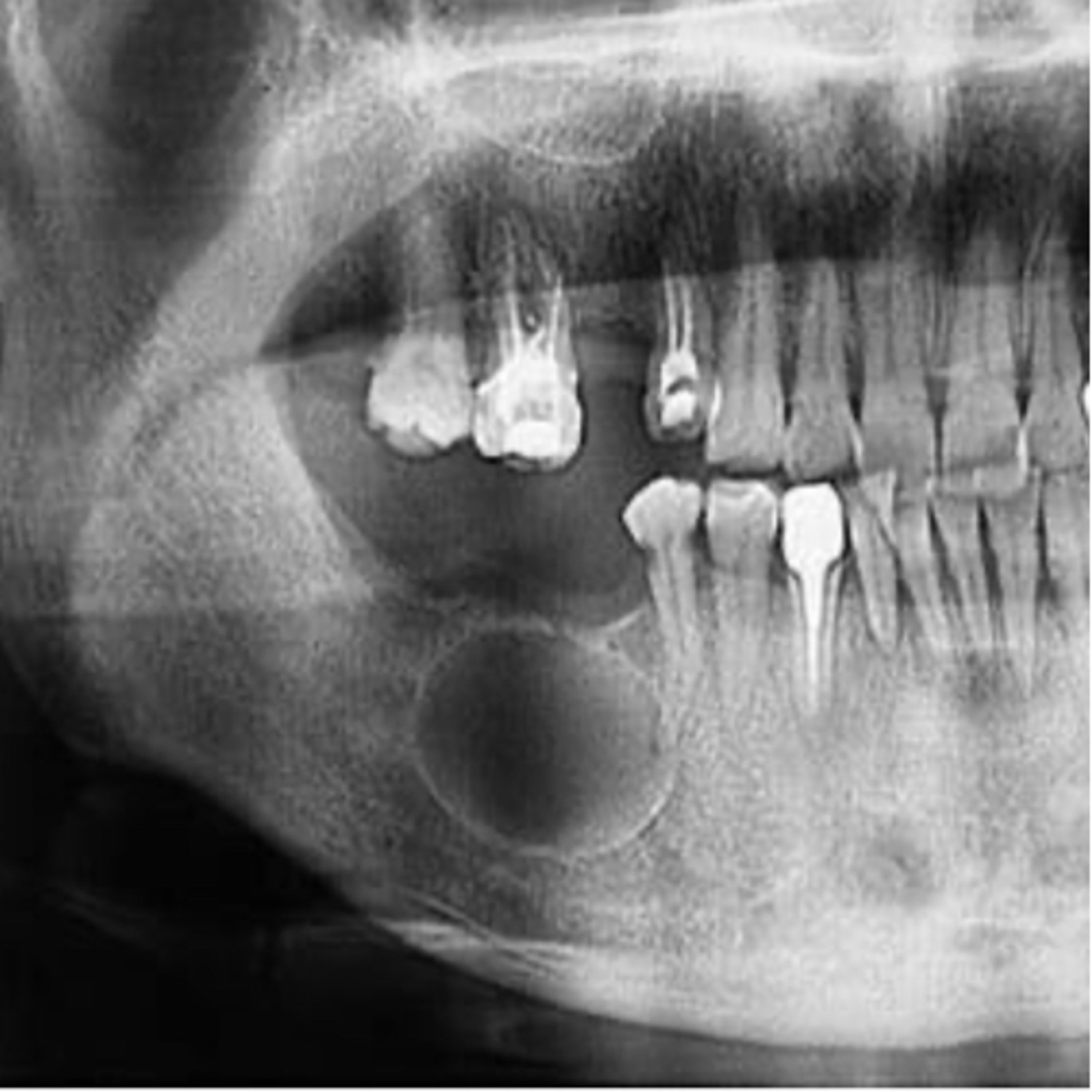
“What are the Differences Between a Primordial Cyst and an OKC?”
7.9.24 Quick Review #172 - #pathology #oralpathology #doctorgallagher #oralsurgery #oralsurgeon #dentist #dentistry #dental Clinically: Primordial Cyst: • Rarely diagnosed clinically because they are often discovered incidentally on radiographs. • Generally asymptomatic unless secondarily infected. • Found in areas where a tooth failed to develop, often in the third molar region. Odontogenic Keratocyst (OKC): • May present with swelling, pain, or drainage if infected. • Can cause displacement of teeth or resorption of adjacent bone. • Often associated with a high recurrence rate and may require long-term follow-up. • Frequently seen in the posterior mandible. Radiographically: Primordial Cyst: • Appears as a well-defined, unilocular radiolucency in the area of a missing tooth. • The absence of the involved tooth is a key radiographic feature. Odontogenic Keratocyst (OKC): • Can appear as unilocular or multilocular radiolucencies. • Well-defined, often with scalloped borders. • Tendency to grow in an anteroposterior direction without causing significant bone expansion. • May be associated with unerupted teeth. Histopathologically: Primordial Cyst: • Initially considered to be a cyst that arises from the degeneration of the dental lamina before any tooth development. • Histopathologically, the lining is similar to that of a dental follicle or reduced enamel epithelium. Odontogenic Keratocyst (OKC): • Characterized by a parakeratinized stratified squamous epithelium lining. • The epithelial lining is typically 6-10 cell layers thick. • Basal cells show a palisaded arrangement with a hyperchromatic appearance. • Presence of a corrugated surface layer of parakeratin. • May exhibit satellite cysts or daughter cysts in the cyst wall. • High mitotic activity and epithelial proliferation are often observed, contributing to the aggressive nature and recurrence rate of OKCs. Summary: • Primordial cysts are primarily identified where a tooth did not develop and are histologically similar to a dental follicle or reduced enamel epithelium. • OKCs are more aggressive, have a higher recurrence rate, and are histologically distinct with a parakeratinized stratified squamous epithelium, palisaded basal layer, and potential for satellite cysts. References: 1. Inspired Pencil. (2023). Primordial cyst. Retrieved from https://lnkd.in/eMeAZUQz 2. Neville, B. W., Damm, D. D., Allen, C. M., & Chi, A. C. (2015). Oral and Maxillofacial Pathology (4th ed.). Elsevier. 3. Miloro, M., Ghali, G. E., Larsen, P. E., & Waite, P. (Eds.). (2012). Peterson’s Principles of Oral and Maxillofacial Surgery (3rd ed.). PMPH-USA. 4. Shear, M., & Speight, P. (2007). Cysts of the Oral and Maxillofacial Regions (4th ed.). Wiley-Blackwell. 5. ChatGPT. 2024. #podcast #podcasts #dentalpodcast #dentalpodcasts #doctorgallagherpodcast #doctorgallagherspodcast #doctor #dentistry #oralsurgery #dental #viral #dentalschool #dentalstudent #omfs #surgeon #doctorlife #dentistlife #residency #oralsurgeon #dentist #doctorgallagher
You May Also Like

Healing TRAUMA
Kimberly's Hope LLC

人生を自分仕様に整えるラジオ 〜誰かの正解より、自分の納得を大切に〜
寅吉

Spartan Discipline Daily | No-Excuses Mental Toughness Boosts
Peak State Labs

THE DR. BETH'S podcast
Dr. Beth

ADHD, But Make It Spiritual
Peak State Labs

Thrasherway Podcast
HLT

A2 | Dutch Short Stories: Reddit
Dutch Fluency
Kabbalah Media | mp3 #kab_por
Bnei Baruch Kabbalah Education and Research Institute, Бней Барух – Ассоциация "Каббала Ла-Ам"
Kabbalah Media | mp3 #kab_fre
Bnei Baruch Kabbalah Education and Research Institute, Бней Барух – Ассоциация "Каббала Ла-Ам"
Kabbalah Media | mp3 #kab_ita
Bnei Baruch Kabbalah Education and Research Institute, Бней Барух – Ассоциация "Каббала Ла-Ам"
Healing TRAUMA
Kimberly's Hope LLC
人生を自分仕様に整えるラジオ 〜誰かの正解より、自分の納得を大切に〜
寅吉
Spartan Discipline Daily | No-Excuses Mental Toughness Boosts
Peak State Labs
THE DR. BETH'S podcast
Dr. Beth
ADHD, But Make It Spiritual
Peak State Labs
Thrasherway Podcast
HLT
A2 | Dutch Short Stories: Reddit
Dutch Fluency
Kabbalah Media | mp3 #kab_por
Bnei Baruch Kabbalah Education and Research Institute, Бней Барух – Ассоциация "Каббала Ла-Ам"
Kabbalah Media | mp3 #kab_fre
Bnei Baruch Kabbalah Education and Research Institute, Бней Барух – Ассоциация "Каббала Ла-Ам"
Kabbalah Media | mp3 #kab_ita
Bnei Baruch Kabbalah Education and Research Institute, Бней Барух – Ассоциация "Каббала Ла-Ам"
Community discussion