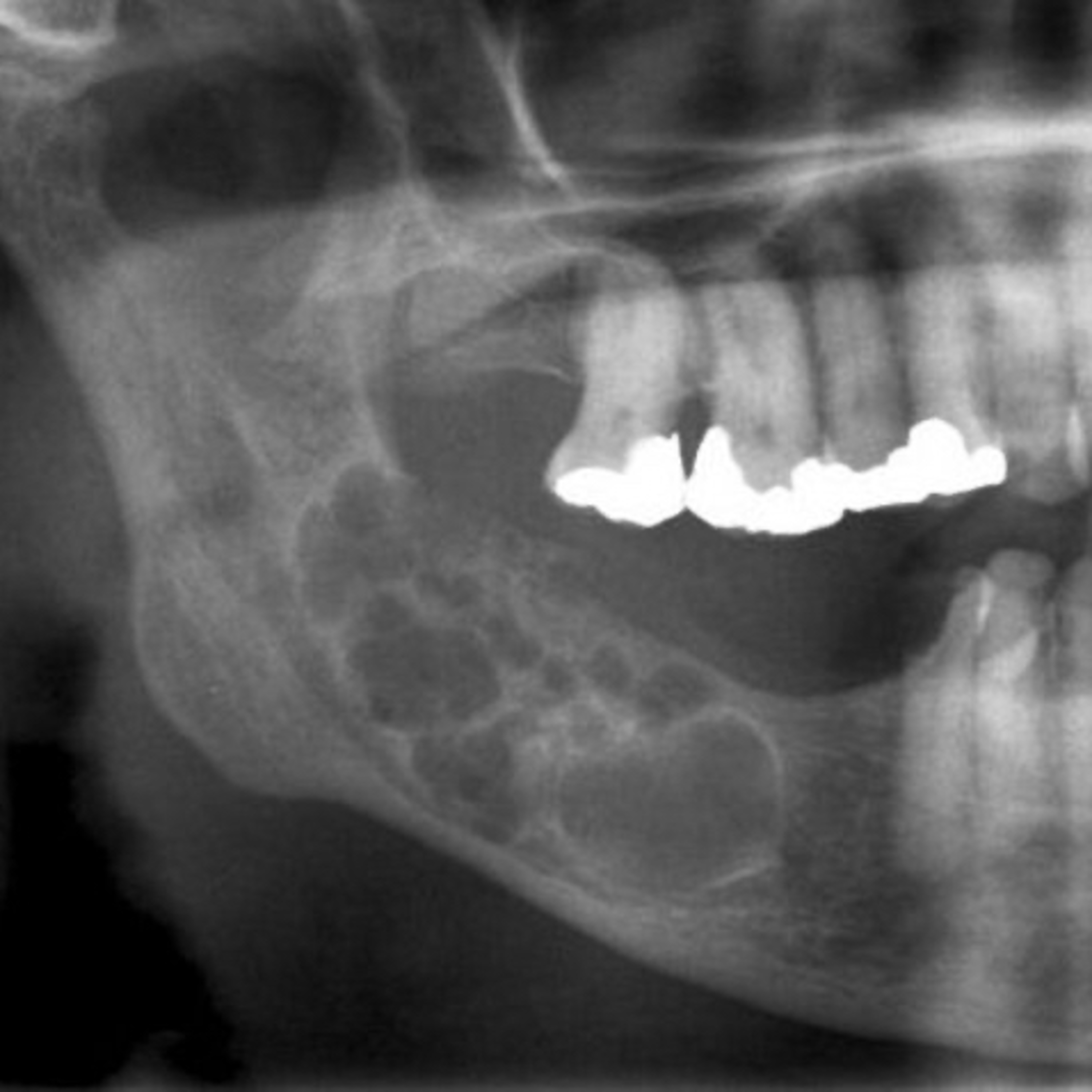
“What are the 6 Subtypes of Conventional (Solid or Multicystic) Ameloblastoma?”
2.16.24 Quick Review #109 - #pathology #oralpathology #doctorgallagher #oralsurgery #oralsurgeon #dentist #dentistry #dental Ameloblastoma, a benign but locally aggressive odontogenic tumor, exhibits 6 subtypes within its most common form, the Conventional (solid or multicystic) Ameloblastoma: 1. Follicular Ameloblastoma: - *Most Common Subtype* - Clinical: Often presents in the posterior mandible as a painless swelling. - Radiographic: Shows well-demarcated, unilocular or multilocular radiolucencies often described as "soap bubble" or "honeycomb" appearances. - Histological: Follicles or islands of odontogenic epithelium surrounded by a fibrous stroma. Central stellate reticulum-like cells with peripheral columnar or cuboidal ameloblast-like cells. 2. Plexiform Ameloblastoma: - Clinical: Similar to the follicular subtype, with a preference for the mandible. - Radiographic: Radiolucent, often multilocular lesions, but the internal structure may appear more "net-like" due to the plexiform pattern. - Histological: Anastomosing strands or cords of odontogenic epithelium within the stroma, resembling a plexus. 3. Acanthomatous Ameloblastoma: - Clinical: Slightly more aggressive behavior and may cause more bone destruction. - Radiographic: Radiolucent areas, similar to other subtypes. - Histological: Notable for squamous metaplasia and keratin formation within the epithelial islands, resembling squamous epithelium. 4. Granular Cell Ameloblastoma: - Clinical: Tends to occur in a slightly older age group, with a slow-growing mass in the jaw. - Radiographic: Radiolucent areas, similar to other subtypes. - Histological: Granular eosinophilic cytoplasm of the stellate reticulum-like cells, giving a granular appearance. 5. Desmoplastic Ameloblastoma: - Clinical: Often involves the anterior maxilla; less common for other subtypes. - Radiographic: Distinct for its mixed radiolucent-radiopaque "honeycomb" or "soap bubble" appearance, often mistaken for fibro-osseous lesions. - Histological: Extensive stromal collagenization (desmoplasia) with small, compressed islands of odontogenic epithelium. 6. Basal Cell Ameloblastoma: - *Least Common Subtype* - Clinical: Rare and may present similarly to other ameloblastomas; can have a more aggressive course. - Radiographic: Radiolucent areas, similar to other subtypes. - Histological: Uniform, small, basaloid cells with less cytoplasm, arranged in compact islands with peripheral palisading. References: 1. Reichart, P. A., Philipsen, H. P., & Sonner, S. (2000). Ameloblastoma: biological profile of 3677 cases. European Journal of Cancer Part B: Oral Oncology, 36(1), 46-50. https://lnkd.in/dg6m2eiq 2. Neville, B. W., Damm, D. D., Allen, C. M., & Chi, A. C. (2015). Oral and Maxillofacial Pathology (4th ed.). Saunders. 3. Abubaker, A.O., Lam, D., & Benson, K. (2016). OMFS Secrets (3rd ed.). Elsevier. 4. ChatGPT. 2024.
You May Also Like

Chat GPT Questions
Sol Good Network

Chat GPT Collective Consciousness
Sol Good Network

Fluent Fiction - Korean
FluentFiction.org

FluentFiction - Afrikaans
FluentFiction.org

Fluent Fiction - Norwegian
FluentFiction.org

Fluent Fiction - Russian
FluentFiction.org

FluentFiction - Finnish
FluentFiction.org

FluentFiction - Czech
FluentFiction.org

FluentFiction - Romanian
FluentFiction.org

Fluent Fiction - Arabic
FluentFiction.org
Chat GPT Questions
Sol Good Network
Chat GPT Collective Consciousness
Sol Good Network
Fluent Fiction - Korean
FluentFiction.org
FluentFiction - Afrikaans
FluentFiction.org
Fluent Fiction - Norwegian
FluentFiction.org
Fluent Fiction - Russian
FluentFiction.org
FluentFiction - Finnish
FluentFiction.org
FluentFiction - Czech
FluentFiction.org
FluentFiction - Romanian
FluentFiction.org
Fluent Fiction - Arabic
FluentFiction.org
Community discussion