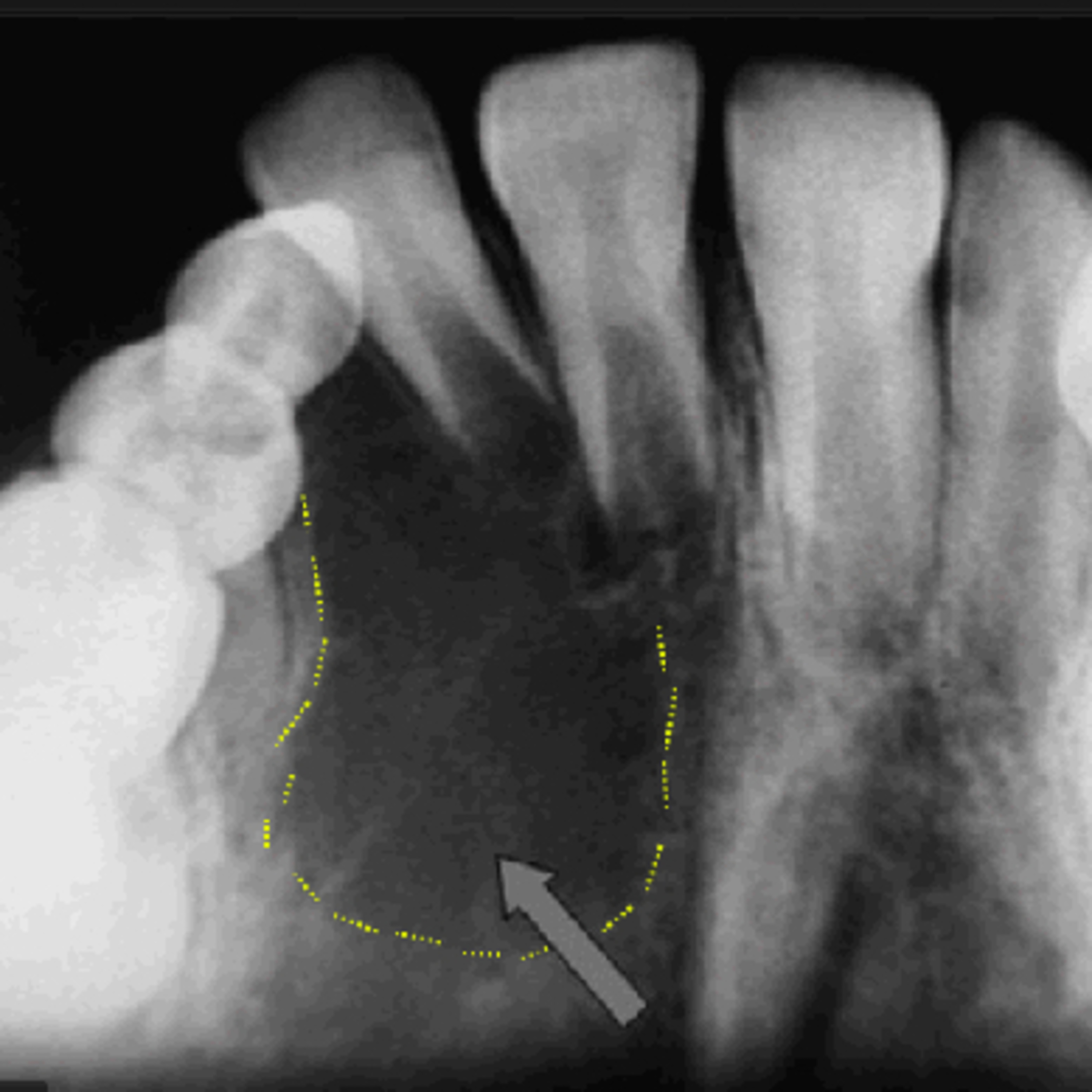
“How Does The Aneurysmal Bone Cyst Differ From Similar Lesions?”
4.23.24 #podcast #podcasts #dentalpodcast #dentalpodcasts #doctorgallagherpodcast #doctorgallagherspodcast Quick Review #127 - #pathology #oralpathology #doctorgallagher #oralsurgery #oralsurgeon #dentist #dentistry #dental Aneurysmal Bone Cyst (ABC) • Clinical Characteristics: - Occurs predominantly in children and young adults. - Rapid, painless swelling; may become painful if there is impingement on nerves or other structures. - Commonly affects the long bones and vertebrae, but also seen in the jaws. • Radiographic Findings: - Exhibits a "soap bubble" or "honeycomb" appearance, radiolucent. - Expansile, multilocular lesion with well-defined borders. - Can cause thinning of the cortex, showing ballooning of the bone. • Histological Features: - Multiple blood-filled cystic spaces without endothelial lining. - Fibrous septa containing multinucleated giant cells, osteoid, and reactive woven bone. - Occasionally shows areas of solid fibrous proliferation. Differential Diagnoses and Differentiating Features: 1. Ameloblastoma - Clinical: Typically slower growing; occurs mainly in adults. - Radiographic: “Honeycombed" or "soap bubble" radiolucencies, but more localized and defined; does not typically cause the same degree of expansile distortion. - Histological: Consists of odontogenic epithelial cells arranged in palisaded columns with reverse polarity; lacks blood-filled cystic spaces. 2. Odontogenic Keratocyst (OKC) - Clinical: Can be aggressive; high recurrence rate. - Radiographic: Presents as a unilocular or multilocular radiolucency; scalloping around the roots of teeth. - Histological: Lined by thin, parakeratinized epithelium; contains keratin debris; no giant cells or blood-filled spaces. 3. Traumatic Bone Cyst (TBC) - Clinical: Often asymptomatic; discovered incidentally. - Radiographic: A unilocular radiolucency without a sclerotic border, often scalloping between the roots. - Histological: An empty cavity or one with serous fluid; lacks an epithelial or cellular lining; no solid tissue components. 4. Giant Cell Granuloma (GCG) - Clinical: More common in younger individuals; may present with pain or swelling. - Radiographic: Radiolucent lesion, can be unilocular or multilocular; lacks the expansile feature seen in ABCs. - Histological: Contains multinucleated giant cells in a background of fibrous stroma; reactive bone present but lacks blood-filled spaces typical of ABC. References: 1. Exodontia.info. (n.d.). Aneurysmal Bone Cyst. Retrieved April 22, 2024, from https://lnkd.in/dH835BTA 2. Motamedi, M. H. K., & Yazdi, E. (2012). Aneurysmal bone cyst of the jaws: Analysis of 11 cases. Journal of Oral and Maxillofacial Surgery, Medicine, and Pathology, 24(3), 175-178. doi:10.1016/j.ajoms.2011.08.002 3. Neville, B. W., Damm, D. D., Allen, C. M., & Bouquot, J. E. (2016). Oral and Maxillofacial Pathology (4th ed.). St. Louis, MO: Elsevier. 4. ChatGPT. 2024.
You May Also Like

Motivational Speeches
Motivationly

Hamza Selfimprovement
Adonis Gang

Playlist Culture G : les podcasts pour apprendre chaque jour
Acast France

Life Hacks DIY & More - Transform Your Everyday With Simple Tricks and DIY Magic!
Byte & Pieces

Happy English Podcast
Michael DiGiacomo Happy English

Un bonbon sur la langue
RTL

Common Law
Flavius Josephus

Neville Goddard - Classic Radio
Neville Goddard

Neville Goddard - Old Time Radio
Neville Goddard

Neville Goddard Daily
Neville Goddard
Motivational Speeches
Motivationly
Hamza Selfimprovement
Adonis Gang
Playlist Culture G : les podcasts pour apprendre chaque jour
Acast France
Life Hacks DIY & More - Transform Your Everyday With Simple Tricks and DIY Magic!
Byte & Pieces
Happy English Podcast
Michael DiGiacomo Happy English
Un bonbon sur la langue
RTL
Common Law
Flavius Josephus
Neville Goddard - Classic Radio
Neville Goddard
Neville Goddard - Old Time Radio
Neville Goddard
Neville Goddard Daily
Neville Goddard
Community discussion